
Crayfish Neural Activity
Measuring Action Potentials in Crayfish
Measuring Neuron Activity
This project explored synaptic connectivity in the crayfish neuromuscular system by correlating presynaptic action potentials from ganglionic nerve 3 with postsynaptic responses in the superficial flexor muscle. The goal was to characterize EPSPs and identify patterns of selective innervation and synaptic summation.
I conducted the background research to inform the design, then performed the dissection, positioned and calibrated extracellular and intracellular electrodes, and collected synchronized voltage recordings under spontaneous and stimulated conditions. The resulting data allowed for classification of action potentials and analysis of corresponding EPSPs and synaptic integration.
The Setup
The crayfish was anesthetized on ice, and the tail was dissected to expose the superficial flexor muscle while preserving ganglionic nerve 3. The preparation was secured in chilled saline to maintain stability and viability.
A suction electrode recorded presynaptic action potentials from the nerve, while an intracellular microelectrode was inserted into individual muscle fibers, confirmed by a stable negative resting potential.
Once calibrated, synchronized extracellular and intracellular recordings were collected under spontaneous and stimulated conditions for analysis.
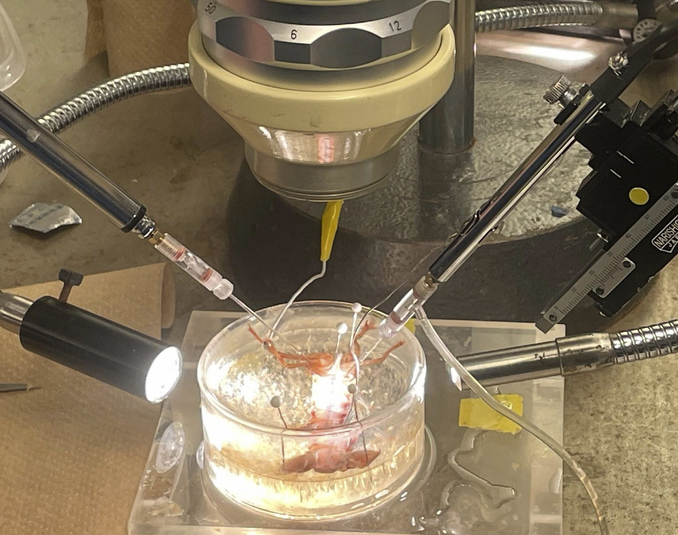
Pinned crayfish tail with attached extracellular electrode (right) and intracellular electrode (left)
Results
The synchronized extracellular and intracellular recordings were analyzed in MATLAB to quantify EPSP amplitude, rise time, and duration and to correlate postsynaptic responses with presynaptic action potentials. Distinct action potential classes were identified, and EPSPs were matched to the nearest preceding spikes to assess connectivity patterns.
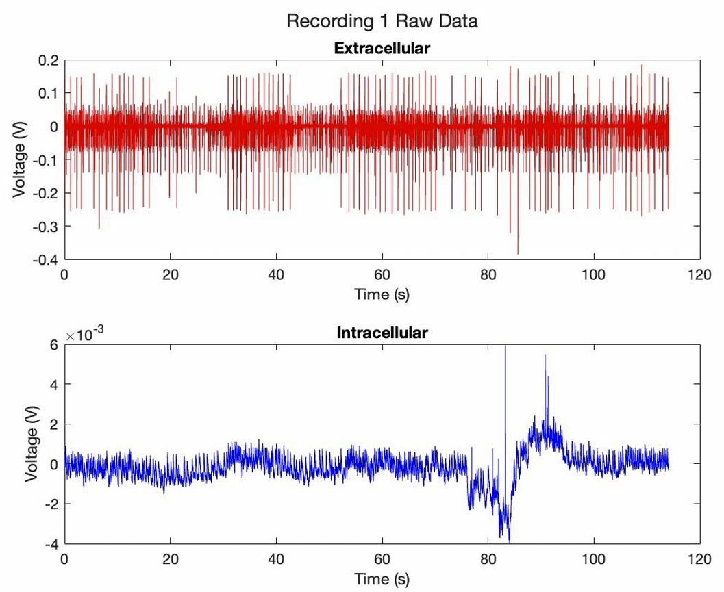
The raw intracellular and extracellular data from the measured recording. Muscle 1 is not inhibited. Muscle 2 is inhibited via tailfan stimulation.
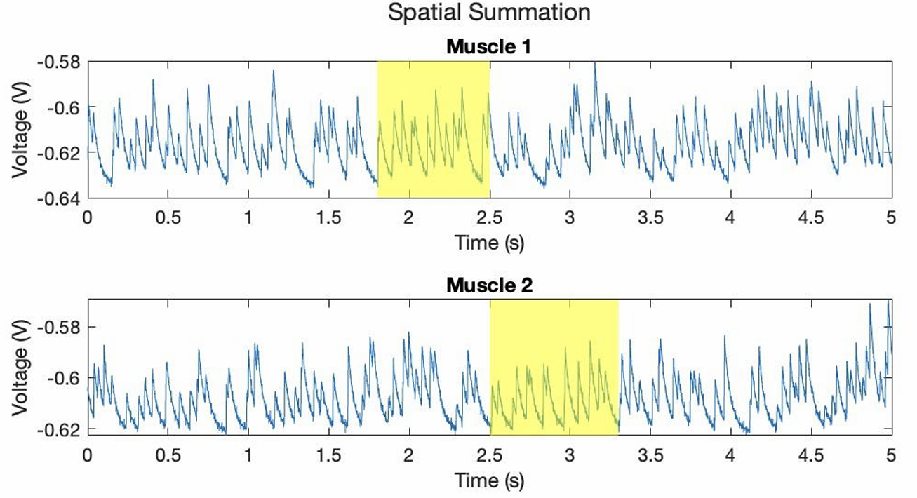
The analysis revealed selective innervation, as not every action potential produced a measurable EPSP, and demonstrated both temporal and spatial summation within individual muscle fibers. Inhibition reduced EPSP amplitude and altered kinetics, highlighting modulation within the system.
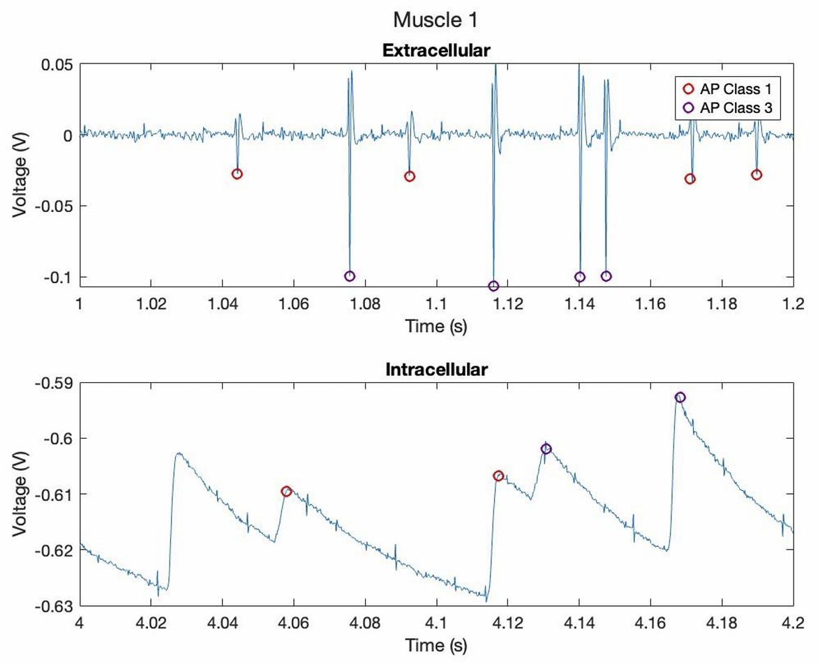
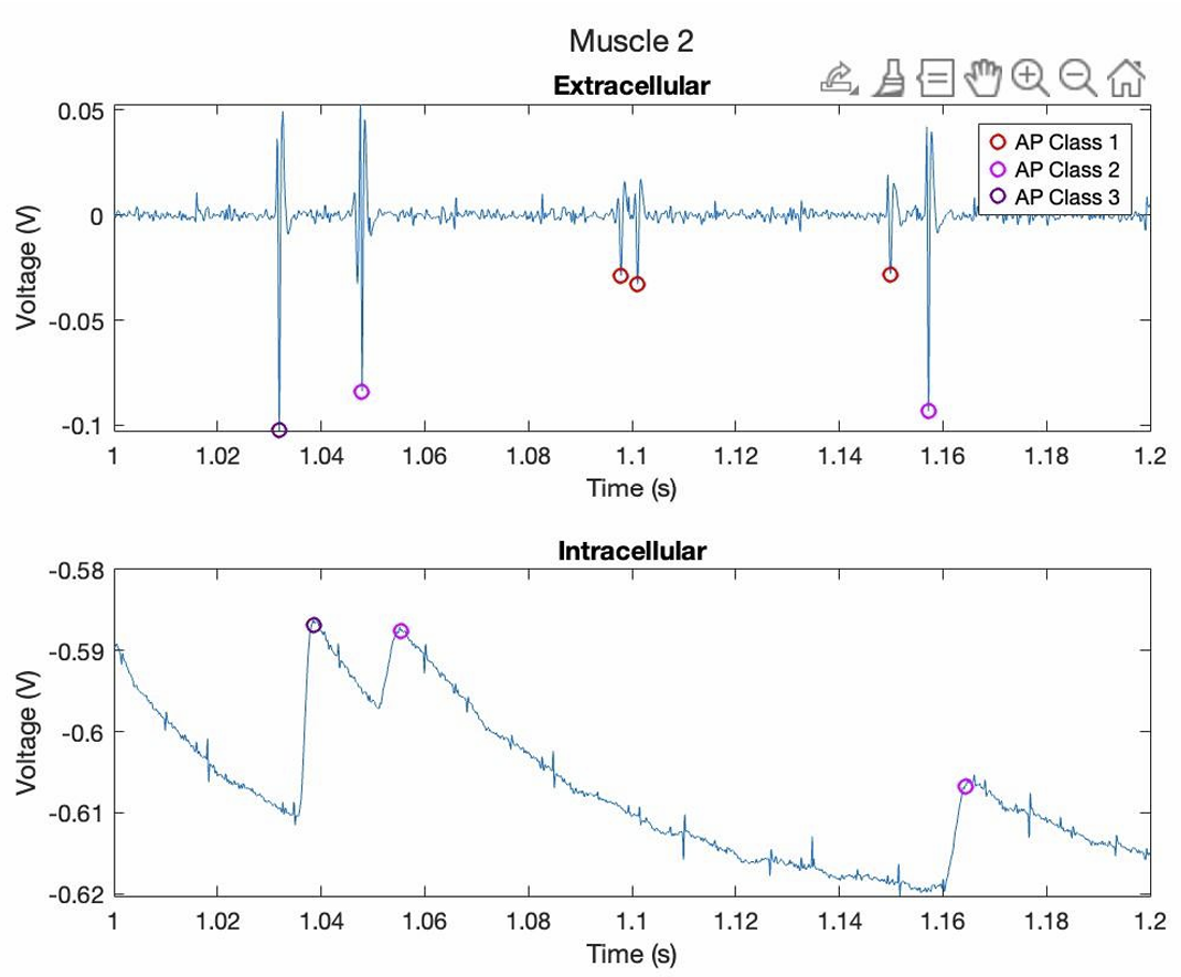
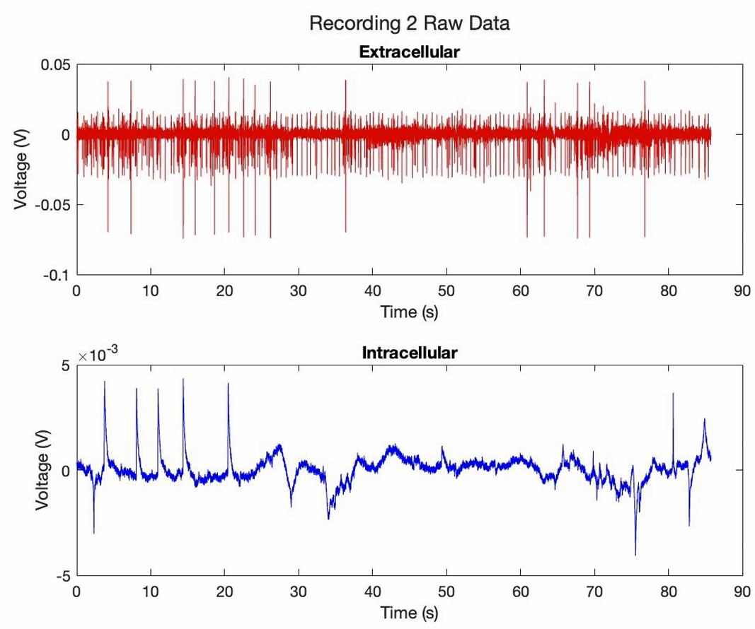
Action potential and EPSP data for muscles one & two. Muscle one showed EPSPs for both action‑potential classes, while muscle two showed EPSPs only for classes 2 and 3, not class 1.
Overall, the results confirmed that the crayfish neuromuscular junction models graded synaptic transmission, selective connectivity, and synaptic integration—core principles of more complex neural systems.
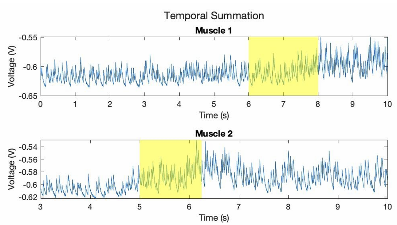
Spatial & temporal summation in muscles one and two.